
Fine Needle Aspiration in Pets: A Clear Guide for Owners
When a dog develops a lump, bump, or swelling, it can be alarming. While not every mass is dangerous, determining what’s going on beneath the surface is essential for your dog’s health and your peace of mind. One of the most commonly used, minimally invasive diagnostic tools in veterinary medicine is fine needle aspiration (FNA). This simple procedure helps veterinarians gather valuable information about abnormal tissues without the need for surgery (Griffiths et al., 1984).
In this article, we’ll explore what fine needle aspiration is, how it works, when it’s recommended, and what pet owners can expect before, during, and after the procedure.
What Is Fine Needle Aspiration?
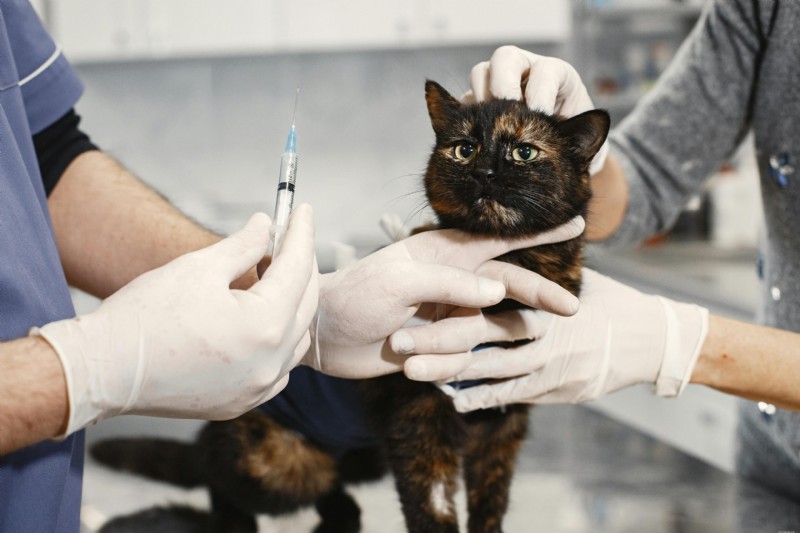
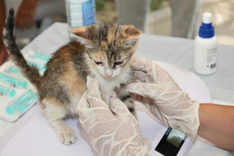
Fine needle aspiration is a diagnostic procedure in which a veterinarian uses a thin, hollow needle to collect a cell sample, often consisting of individual cells or small clusters, from a mass or abnormal area on a dog’s body. The collected cells are then placed on a microscope slide, stained, and examined either in-house or by a veterinary pathologist.
Fine needle aspiration is also known as a fine needle biopsy. Unlike a traditional biopsy, which involves removing a piece of tissue, FNA collects only individual or small clusters of cells. Because of this, it is less invasive, quicker, and typically does not require anesthesia.
FNA is primarily a diagnostic tool to determine if a mass is cancerous, noncancerous, or related to an infection.
Why Is Fine Needle Aspiration Used?
Dogs can develop lumps for many reasons, some harmless, others more serious. Fine needle aspiration helps veterinarians distinguish between different types of masses, including suspicious lumps, such as:
-
Lipomas (fatty tumors) – usually benign and common in older dogs
-
Cysts – fluid-filled sacs that may or may not require treatment; FNA can be used to obtain a fluid sample for diagnosis or to therapeutically drain fluid, providing relief from discomfort and preventing complications
-
Abscesses – pockets of infection that need drainage and antibiotics; FNA can help obtain a fluid sample to guide treatment
-
Inflammatory lesions – caused by injury or infection
-
Tumors (benign or malignant) – including mast cell tumors or carcinomas
-
FNA is commonly used to biopsy newly identified masses in the breast, thyroid, lymph nodes, and other areas of the body
The main goal of FNA is to determine the nature of the mass so that an appropriate treatment plan can be developed.
When Is FNA Recommended?
Veterinarians often recommend fine needle aspiration whenever a new lump or swelling is discovered. Even if a mass appears harmless, visual inspection alone cannot reliably determine its nature.
Some specific situations where FNA is commonly used include:
-
A newly discovered lump under the skin
-
A mass that is growing or changing shape
-
Swollen or enlarged lymph nodes
-
Thyroid nodules
-
Abnormal organs detected through imaging (such as ultrasound guidance)
-
Persistent skin lesions or sores
FNA uses real-time guidance, often with ultrasound, to precisely target lesions. It can be performed on almost any region of the body, but is particularly effective for assessing masses in the pancreas, gastrointestinal tract, and mediastinum when guided by imaging techniques such as ultrasound or CT scans.
Early evaluation is critical. Waiting to see if a lump changes can delay diagnosis and treatment, especially if the mass turns out to be malignant.
Preparation and Planning
Before a fine needle aspiration (FNA) procedure, careful preparation is essential to ensure the safety and comfort of your dog. The process begins with your veterinarian reviewing your pet’s medical history, including any current medications and known allergies. This step helps identify any factors that could increase the risk of bleeding complications, such as the use of blood thinners or underlying health conditions.
Your veterinarian will explain the FNA procedure, outlining its benefits, potential risks, and what to expect during and after the needle aspiration. This is also the time to ask any questions or share concerns you may have about the biopsy site or the process itself. In some cases, you may be advised to temporarily stop certain medications to minimize the risk of bleeding.
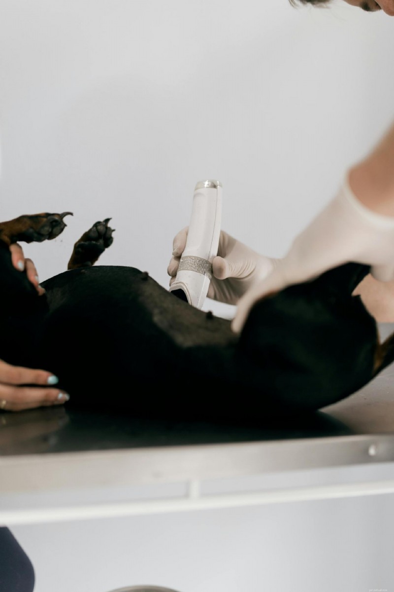
On the day of the procedure, the area where the fine needle will be inserted is cleaned and disinfected to reduce the risk of infection. For most dogs, a local anesthetic is applied to numb the biopsy site and ensure the FNA procedure is as comfortable as possible. If the mass is located deep within the body (such as in the abdominal cavity or within deep seated lesions) imaging tests like ultrasound or CT scans may be used to guide the needle precisely to the abnormal area.
By following these preparation steps, healthcare providers can help ensure that the fine needle aspiration FNA is performed safely and effectively, providing valuable information for diagnosis and treatment planning.
Equipment and Personnel
A successful fine needle aspiration biopsy relies on the right equipment and skilled personnel. The primary tool is a very thin needle, typically ranging from 21 to 30 gauge, which can be attached to a syringe for gentle suction during the needle aspiration. The collected biopsy sample is then placed in a sterile container or directly onto a microscope slide for examination.
For masses that are difficult to locate or are deep within the body, imaging equipment such as ultrasound or CT scanners may be used to guide the fine needle to the precise location. This imaging guidance is especially important for obtaining an accurate diagnosis from deep seated lesions or abnormal tissue that cannot be easily felt.
The FNA procedure is usually performed by a veterinarian or a specialist with experience in fine needle aspiration biopsy, such as a radiologist or pathologist. Additional personnel, including veterinary nurses or technicians, may assist with patient preparation, monitoring, and support throughout the procedure. Maintaining a sterile environment and using proper technique are crucial to minimize the risk of infection and ensure the quality of the biopsy sample, ultimately leading to a more accurate diagnosis (Simeonov, 2012).
How the Procedure Works
Fine needle aspiration is straightforward and typically takes only a few minutes. Here’s what usually happens:
-
Preparation
The area may be cleaned, but shaving is not always necessary. Most dogs tolerate the procedure well without sedation. Local anesthesia may be used depending on the location and sensitivity of the area, especially for deeper or more invasive procedures. -
Sample Collection
A small needle, typically ranging from 21 to 30 gauge (needle size), is inserted into the mass. The veterinarian may apply slight suction with a syringe to collect cells. Multiple samples may be collected from different areas of the mass to improve diagnostic accuracy and ensure representative tissue is obtained. -
Slide Preparation
The material obtained from the mass is placed onto a glass slide and spread thinly. -
Staining and Examination
The slide is stained and examined under a microscope. In some cases, it is sent to a laboratory for further analysis.
The entire FNA process usually takes between 15 and 30 minutes for an outpatient procedure. The process is quick, and many dogs barely notice it happening (Simeonov, 2012).
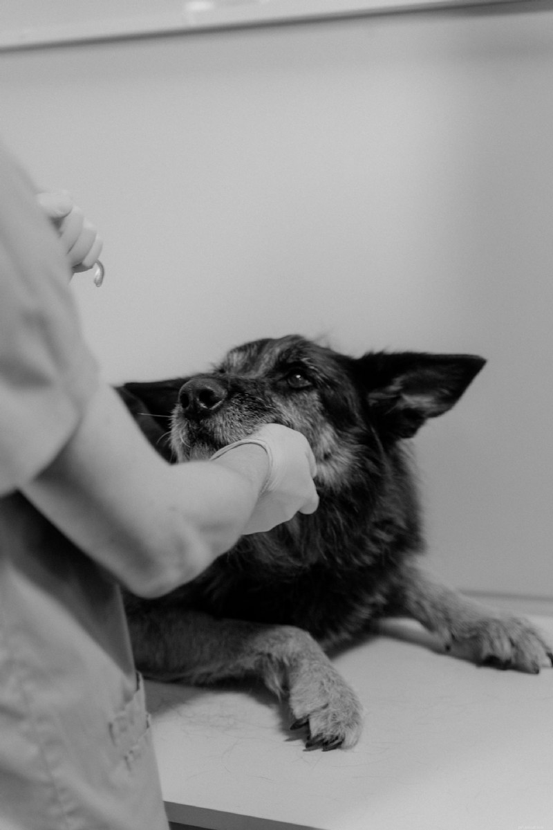
Does FNA Hurt Dogs?
One of the most common concerns pet owners have is whether the procedure is painful. Fortunately, fine needle aspiration is generally well tolerated. The needle used is very small, and most dogs experience little to no discomfort (similar to receiving a vaccine).
In rare cases, if the mass is in a sensitive area or the dog is particularly anxious, mild sedation may be recommended. However, this is not the norm.
Benefits of Fine Needle Aspiration
FNA offers several advantages that make it a preferred first step in evaluating lumps:
-
Minimally invasive – no surgery required; fine needle biopsy and FNA biopsies use a thin needle to collect cells with minimal discomfort
-
High accuracy – studies indicate an overall accuracy rate of 93.1% for FNA in assessing tumors and nodules, supporting a correct diagnosis (Simeonov, 2012)
-
Quick results – sometimes available the same day, and typically within a few days; your healthcare provider will discuss the findings and any necessary follow-up care or additional tests
-
Low cost compared to surgical biopsies
-
No general anesthesia needed in most cases
-
Early detection of potentially serious conditions
-
First-line biopsy choice – FNA is often chosen first due to its speed and minimal discomfort, while a core biopsy may be recommended if FNA results are inconclusive
-
Rapid recovery – after an FNA, a bandage is applied to the site and most patients can return to normal activities immediately
Because it is so simple and accessible, FNA biopsies allow veterinarians to make faster decisions about whether further testing or treatment is necessary, helping to ensure a correct diagnosis and optimal patient outcomes.
Limitations of FNA
While fine needle aspiration is extremely useful, it does have limitations. Because it collects only cells rather than full tissue samples, it may not always provide a definitive diagnosis.
Some potential limitations include:
-
Inconclusive results if not enough cells are collected or if atypical cells are present, which may require molecular testing or further biopsies
-
Difficulty diagnosing certain tumor types
-
Inability to assess tissue architecture, which is sometimes necessary for accurate diagnosis
A core biopsy gathers more tissue than FNA, providing more detailed information about the cell type and structure, which can be crucial for accurate diagnosis. Molecular testing, including genetic and biomarker analysis, can enhance diagnostic accuracy and guide targeted therapies, especially when cytological results are inconclusive or show atypical cells.
If the results are unclear or if a more detailed analysis is needed, your veterinarian may recommend a biopsy or additional tests.

Special Considerations
While fine needle aspiration is a minimally invasive and generally safe procedure, there are special considerations that healthcare providers must keep in mind to ensure the best possible outcome. Patients taking anticoagulant medications or those with a history of bleeding disorders are at a higher risk for bleeding complications during or after the needle aspiration. In these cases, your veterinarian may recommend adjusting medications or taking additional precautions to minimize significant complications (Vos et al., 1989).
In cosmetically sensitive areas, such as the thyroid or other visible locations, fine needle aspiration is often preferred over core needle biopsy or excisional biopsy. The use of a fine needle helps reduce scarring and promotes faster healing, making it an ideal choice for areas where appearance is a concern.
If the FNA reveals abnormal cells or suggests the presence of malignant tumors, further testing may be necessary. This can include molecular markers or genetic analysis to guide cancer treatments and provide a more accurate diagnosis. In some cases, FNA is used to remove fluid from a suspicious mass, and ongoing monitoring may be required to check for recurrence or the need for further evaluation.
After the procedure, your referring physician or veterinarian will receive a detailed report of the biopsy results, including recommendations for any additional testing or treatment. Early detection and accurate diagnosis are key to effective patient care, helping to guide treatment decisions and improve outcomes for your dog. By working closely with your healthcare providers and staying informed, you can help ensure the best possible care for your pet.
Understanding the Results
After the sample is examined, the results generally fall into one of several categories:
-
Benign – non-cancerous and usually not life-threatening; FNA can help identify benign growths and distinguish them from malignant lesions
-
Malignant – cancerous and may require treatment such as surgery, chemotherapy, or radiation; FNA can detect cancer cells and help determine if metastatic disease is present
-
Inflammatory or infectious – indicating infection or immune response
-
Inconclusive – not enough information to make a diagnosis
Additionally, genetic or molecular markers identified in the FNA sample can help guide personalized cancer treatment by predicting responsiveness to specific therapies (Benaduce et al., 2023).
Your veterinarian will explain the findings and discuss the next steps. In some cases, monitoring the mass may be sufficient; in others, immediate treatment may be necessary.
What Happens After the Procedure?
One of the advantages of FNA is that recovery is essentially immediate. Most dogs resume normal activity right away.
You may notice:
-
Slight swelling or redness at the site
-
Minor bleeding (rare and usually minimal)
These effects typically resolve quickly without intervention. If you notice persistent swelling, pain, or changes in your dog’s behavior, it’s a good idea to contact your veterinarian.
When a Biopsy Is Needed
If fine needle aspiration does not provide a clear diagnosis, a biopsy may be recommended. Fine needle aspiration (FNA) is less invasive than a core needle biopsy, which uses a larger needle and may require a small incision to obtain a tissue sample. In some cases, a standard incision may be used for more invasive procedures like incision and drainage or tissue sampling. If biopsy results are inconclusive or if the lesion's nature requires it, surgical excision may be necessary as the definitive treatment for diagnosis or removal.
A biopsy can provide more detailed information, including how aggressive a tumor may be and whether it has invaded surrounding tissues. While more invasive, it is sometimes necessary for accurate diagnosis and treatment planning (Vos et al., 1989).
The Importance of Early Detection
One of the key takeaways for dog owners is the importance of early evaluation. Many serious conditions, including cancer, are far more treatable when caught early. Using fine needle aspiration to biopsy newly identified masses can improve outcomes and facilitate early treatment.
A good rule of thumb is:
-
Check your dog regularly for new lumps or changes
-
Measure and monitor any existing masses
-
Schedule a veterinary visit if anything changes
Fine needle aspiration makes it easy to investigate concerns quickly and with minimal stress for your pet.
Common Myths About FNA
There are a few misconceptions about fine needle aspiration that can cause unnecessary worry:
Sticking a needle into a tumor can spread cancer
This is a common myth. In reality, the risk of spreading cancer through FNA is extremely low and not considered a significant concern in veterinary medicine. FNA is very safe, with a reported morbidity rate of only 0.98% (Dank et al., 2025). However, there are rare risks such as bleeding, infection (especially if performed in an infected area), damage to surrounding structures, and needle tract seeding, where tumor cells may be deposited along the needle tract. Patients who are coagulopathic or taking anticoagulant medications may have an increased risk of bleeding complications, but the incidence remains low.
If the lump isn’t bothering my dog, it’s fine
Many serious conditions do not cause pain initially. Lack of discomfort does not mean a mass is harmless.
All lumps in older dogs are fatty tumors
While lipomas are common, not all lumps are benign. Testing is the only way to be sure.
Final Thoughts
Fine needle aspiration is a simple yet powerful diagnostic tool that plays a crucial role in modern veterinary care. FNA can be performed with imaging guidance, such as ultrasound imaging, CT guidance, or endoscopic ultrasound, to obtain a tissue or fluid sample from deep or hard-to-reach areas. Using an imaging test helps guide the needle precisely to the correct location, improving accuracy and diagnostic yield. FNA is especially important for diagnosing conditions such as thyroid cancer and metastatic disease, as well as evaluating other abnormal masses to help guide decisions about treatment and monitoring.
For dog owners, the procedure offers reassurance and clarity without putting their pets through unnecessary stress or invasive surgery. If you ever find a lump on your dog, don’t ignore it—having it evaluated early with a procedure like FNA can make all the difference.
By staying proactive and informed, you can help ensure your dog receives the best possible care and maintains a healthy, happy life.
References
Benaduce Emanuelli Mello, C., Engelmann, A. M., Kommers, G. D., Martins Flores, M., Almeida Fighera, R., Rodrigues e Rodrigues, B., ... & Melazzo de Andrade, C. (2023). Fine needle aspiration cytology: High accuracy in diagnosing cutaneous and subcutaneous neoplasms in dogs. Comparative Clinical Pathology, 32(1), 155-164.
Dank, G., Buber, T., Levy-Hirsch, L., Fink, A., Marks, N., & Hanael, E. (2025). Underutilization of Fine Needle Aspiration for Canine and Feline Superficial Masses-Survey Results.
Griffiths, G. L., Lumsden, J. H., & Valli, V. E. O. (1984). Fine needle aspiration cytology and histologic correlation in canine tumors. Veterinary Clinical Pathology, 13(1), 13-17.
Simeonov, R. S. (2012). The accuracy of fine-needle aspiration cytology in the diagnosis of canine skin and subcutaneous masses. Comparative Clinical Pathology, 21(2), 143-147.
Vos, J. H., van den Ingh, T. S. G. A. M., & Van Mil, F. N. (1989). Non‐exfoliative canine cytology: The value of fine needle aspiration and scraping cytology. Veterinary Quarterly, 11(4), 222-231.
- Pet Behavior
- Pet Breeds
- Pet Names
- Pet Adoption
- Pet Training
- Pet Information
- Pet Health
- Adorable Pets
- Dogs
- Decoding Your Cat’s Tail: 7 Key Positions Every Owner Should Master
- Efficiently Cycle Your Saltwater Aquarium Using Live Rock: A Step‑by‑Step Guide
- Cat Stomatitis: Causes, Symptoms, and Effective Treatments
- Understanding Purple Gums in Dogs: When to Worry
- Mozart's Tale: How a Ragdoll Kitten Became the Heart of a Home
- Giant Schnauzer: Size, Temperament, Lifespan & Puppies – Your Comprehensive Breed Guide
- 9 Must-Read Cookbooks for Safe Homemade Dog Food