
Testicular Cancer in Dogs: Early Signs, Causes, and Effective Treatments
Neutering offers vital health benefits for male dogs, including a significant reduction in the risk of testicular cancer. Early detection of tumors can save lives.
What Is Testicular Cancer?
In dogs, testicular cancer accounts for about 90 % of all male reproductive system cancers. Unneutered males are far more susceptible. While most tumors stay localized, metastasis can reach lymph nodes, liver, lungs, and other organs.
The primary tumor types are:
- Leydig Cell Tumors: Originate from testosterone‑producing cells. They are usually small, rarely metastasize, and often remain asymptomatic, so routine exams are key to detection.
- Sertoli Cell Tumors: Develop from sperm‑producing cells, tend to spread, and are more common in undescended testicles. These tumors often show clear signs, making early identification critical.
- Seminomas: Emerge from germ cells in the testicle’s inner layer. Though less aggressive, they can produce specific clinical signs and occasionally coexist with other tumor types.
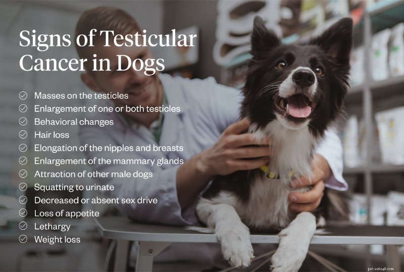
Recognizing the Signs
Signs can vary, so stay alert to any changes:
- Testicle shrinkage or enlargement
- Swelling, discomfort, or skin changes (hair loss, discoloration)
- Suspended foreskin or twisted testicle
- Swollen mammary glands or penile atrophy (feminization)
- Behavioral shifts: lethargy, increased focus on genital area, squatting to urinate
- Loss of appetite or unexplained weight loss
Feminization—caused mainly by Sertoli tumor‑derived estrogen—may manifest as symmetrical hair loss, enlarged nipples, and mandible changes. If you notice these, schedule a vet visit promptly.
Diagnosis & Treatment
Accurate diagnosis guides the best treatment plan. Veterinarians typically employ:
- Physical Examination: Assess size, shape, and texture of the testicles.
- Fine‑Needle Aspiration Cytology: Screens for metastasis and identifies tumor cell type.
- Complete Blood Count (CBC): Detects anemia, low platelets, or leukopenia related to estrogen effects.
- Abdominal Ultrasound: Reveals undescended testicles and regional lymph node status.
- Testicular Ultrasonography: Differentiates malignant from benign lesions with detailed imaging.
Once the tumor type and stage are confirmed, the mandatory step is an orchiectomy—complete removal of the affected testicle. This not only eradicates cancerous cells but also prevents future complications.
In bilateral cases, a bilateral orchiectomy is advised to ensure all malignant tissues are eliminated. For aggressive or metastatic tumors, adjunct therapies such as chemotherapy or radiation may be recommended, especially for seminomas.
Post‑treatment care involves regular follow‑up appointments to monitor recovery and detect any recurrence. For advanced disease, palliative care focuses on improving the dog’s quality of life.
When performed early, surgical removal offers an excellent prognosis—many dogs return to a happy, healthy life.

Proactive veterinary check‑ups and early detection are the cornerstones of successful treatment. If you suspect testicular cancer, collaborate closely with your vet and adhere to recommended follow‑ups.
For convenient, expert care, consider Dutch—a trusted online veterinary service that allows you to consult with specialists from home. Schedule a virtual appointment to discuss your dog’s needs and secure the best possible care.
- Pet Behavior
- Pet Breeds
- Pet Names
- Pet Adoption
- Pet Training
- Pet Information
- Pet Health
- Adorable Pets
- Dogs
- Cat Pancreatitis: The Hidden Threat – Symptoms, Diagnosis & Prevention
- Understanding Dog Eczema: Causes, Symptoms & Effective Treatment
- Do Dogs Sense the Loss of a Family Member?
- Effective Ways to Eliminate Dog Urine Odor in Your Home
- Take a Break: Adorable Raccoons to Brighten Your Day
- Celebrating 15 Years of Caymus: A Heartfelt Birthday Tribute
- Essential Post‑Hurricane Safety: What to Do When Floodwater Rises