

Mast Cell Tumors in Dogs: A Complete Guide to Diagnosis, Treatment & Prognosis
Skin lumps in dogs are often harmless, but some require immediate veterinary attention. Mast cell tumors (MCT) are the most common malignant skin cancer in dogs and can mimic benign growths.
Because they can appear as anything from a tiny soft bump to a large ulcerated mass, MCTs are called “the great pretenders.” Their appearance alone does not reveal how aggressive they are (Misdorp, 2004). Early detection and treatment have dramatically improved outcomes.
This guide covers risk factors, clinical signs, diagnostic methods, staging, treatment options, and what owners can expect when their dog is diagnosed with an MCT.
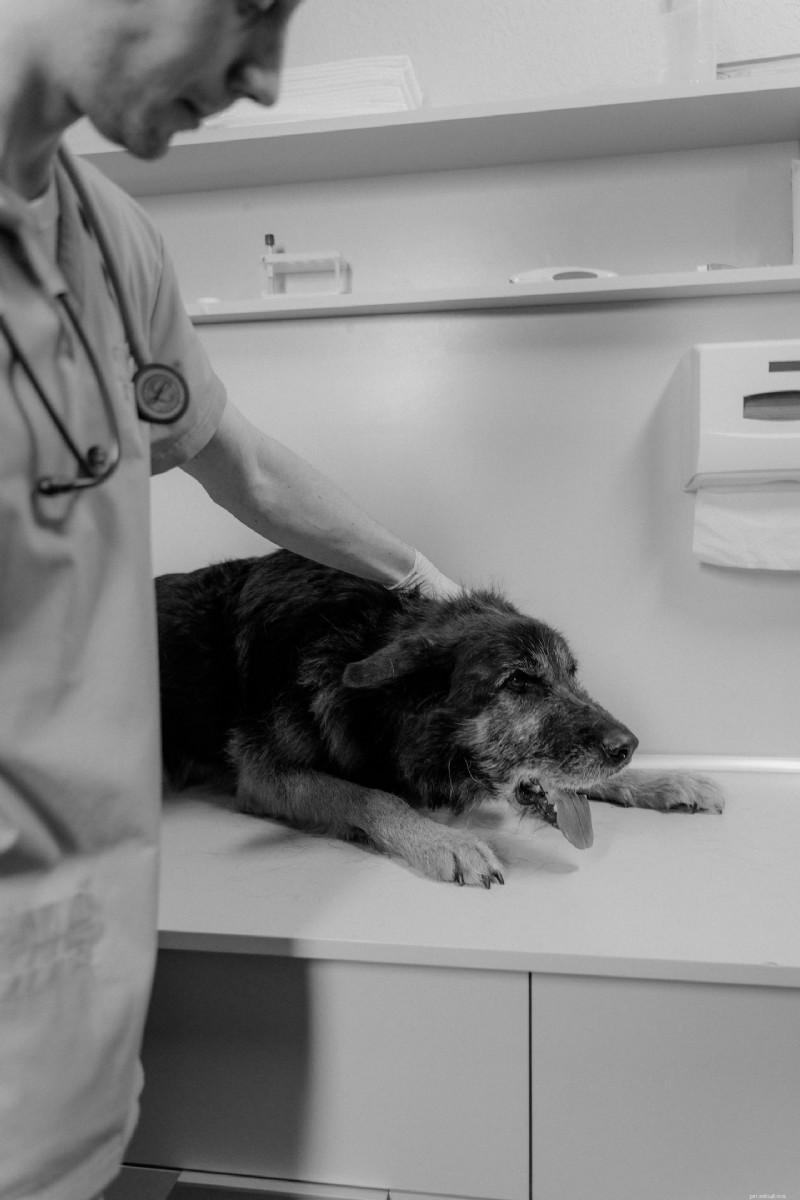
What Are Mast Cell Tumors?
MCTs arise from mast cells—white blood cells that store histamine, heparin, and other inflammatory mediators. Normally they help defend against parasites and trigger allergic reactions. When these cells proliferate uncontrollably, they form tumors that can invade local tissue and, in advanced cases, spread to lymph nodes, spleen, liver, bone marrow, or other organs.
Canine MCTs represent about 16‑21% of all skin tumors, making them the most frequent cutaneous cancer in dogs.
Why MCTs Are Unique
These tumors can change size dramatically within hours due to histamine release, causing swelling, redness, itching, and even ulceration (Horta et al., 2018). Physical manipulation—such as squeezing or excessive handling—can trigger degranulation, temporarily enlarging the mass.
Risk Factors
Genetics
Breed predisposition is strong. Mutations in the KIT gene are common in aggressive MCTs and are especially frequent in breeds such as Boxers, Shar‑Peis, and Golden Retrievers.
Age
Most tumors develop in dogs 8‑10 years old, but aggressive forms can appear in younger dogs (3‑4 years).
Chronic Inflammation
Ongoing skin irritation may contribute to abnormal mast‑cell activity, although evidence is still emerging.
Environment
No single environmental toxin has been linked to canine MCTs; genetics remains the dominant factor.

Breeds at Higher Risk
Boxers, Labrador Retrievers, Golden Retrievers, Boston Terriers, Pugs, Staffordshire Bull Terriers, English & French Bulldogs, Weimaraners, Rhodesian Ridgebacks, Beagles, Schnauzers, and Shar‑Peis are most commonly affected. Mixed‑breed dogs can develop MCTs as well.
Typical Locations
About 80 % of MCTs are cutaneous, appearing on the legs, chest, neck, flanks, abdomen, face, or tail. Internal MCTs (spleen, liver, gastrointestinal tract, bone marrow) are less common but more serious.
Clinical Signs
- Small button‑like bump
- Hairless raised lump
- Soft swelling under the skin
- Wart‑like growth
- Red, inflamed mass
- Ulcerated lesion
- Rapidly growing nodule
Additional signs may include itching, bleeding, scabbing, pain, hair loss, vomiting, diarrhea, loss of appetite, melena, lethargy, and weight loss—especially with aggressive, histamine‑producing tumors.
Why Prompt Evaluation Matters
Even lesions that look benign can be malignant. A quick, minimally invasive fine‑needle aspiration (FNA) often provides a definitive diagnosis.
Diagnostic Process
1. Fine‑needle aspiration – performed in‑clinic, results in 1‑3 days. 2. If mast cells are identified, further work‑up may include surgical biopsy, histopathology, CBC, chemistry panel, urinalysis, lymph‑node aspirates, abdominal ultrasound, chest radiographs, CT, and bone‑marrow evaluation.

Grading and Staging
Grading assesses how abnormal tumor cells appear under a microscope and predicts behavior. The older Patnaik system (Grades I‑III) is still referenced, but many pathologists now use the Kiupel two‑tier system (low vs. high grade).
Staging determines the extent of spread (regional lymph nodes, lungs, liver, etc.) and guides treatment planning.
Patnaik Grades
- Grade I (low): Well‑differentiated, slow‑growing, rarely metastasizes; surgery often curative.
- Grade II (intermediate): Variable behavior; may need additional testing and close monitoring.
- Grade III (high): Poorly differentiated, aggressive, high metastatic potential; multimodal therapy required.
Treatment Options
- Surgery – Gold standard; aims for clean margins (2‑3 cm historically, smaller margins acceptable for low‑grade tumors).
- Radiation therapy – Used when margins are incomplete or surgery is not feasible.
- Chemotherapy – Vinblastine, prednisone, lomustine (CCNU) for high‑grade, multiple, or metastatic disease.
- Targeted therapy – Tyrosine kinase inhibitors (toceranib/Palladia®, masitinib) for KIT‑mutated tumors.
- Antihistamines & supportive meds – Diphenhydramine, famotidine, omeprazole, sucralfate to control histamine effects and gastrointestinal ulcers.

Prognosis
Prognosis depends on grade, margins, tumor size, lymph‑node involvement, and presence of metastasis. Low‑grade tumors with clean margins often result in long, cancer‑free survival. High‑grade or metastatic disease carries a guarded prognosis, but modern multimodal therapy can still provide months to years of good quality life.
Prevention and Early Detection
There is no proven way to prevent MCTs because genetics play a major role. Owners should perform monthly skin checks, schedule regular veterinary exams, and have any new lump evaluated promptly.
Living with an Affected Dog
Many dogs respond well to treatment and return to their normal activities. Close communication with a veterinarian or veterinary oncologist helps balance aggressive therapy with quality‑of‑life considerations.
Key Takeaway
Never ignore a new lump. Early diagnosis via fine‑needle aspiration and timely treatment offers the best chance for a successful outcome.
References
De Nardi, A. B. et al. (2022). Diagnosis, prognosis and treatment of canine cutaneous and subcutaneous mast cell tumors. Cells, 11(4), 618.
Garrett, L. D. (2014). Canine mast cell tumors: diagnosis, treatment, and prognosis. Veterinary Medicine: Research and Reports, 49‑58.
Hahn, K. A. et al. (2008). Masitinib is safe and effective for the treatment of canine mast cell tumors. J. Vet. Intern. Med., 22(6), 1301‑1309.
Horta, R. S. et al. (2018). Assessment of canine mast cell tumor mortality risk. Veterinary Pathology, 55(2), 212‑223.
Misdorp, W. (2004). Mast cells and canine mast cell tumours. Veterinary Quarterly, 26(4), 156‑169.
Romansik, E. M. et al. (2007). Mitotic index is predictive for survival for canine cutaneous mast cell tumors. Veterinary Pathology, 44(3), 335‑341.
Sledge, D. G. et al. (2016). Canine cutaneous mast cell tumors: a combined clinical and pathologic approach. The Veterinary Journal, 215, 43‑54.
Thompson, J. J. et al. (2011). Canine subcutaneous mast cell tumor: characterization and prognostic indices. Veterinary Pathology, 48(1), 156‑168.
- Pet Behavior
- Pet Breeds
- Pet Names
- Pet Adoption
- Pet Training
- Pet Information
- Pet Health
- Adorable Pets
- Dogs
- The Ultimate Guide to Choosing the Best Outdoor Guinea Pig Run
- Top 10 Proven Weight‑Loss Strategies for Senior Dogs
- Compassionate End-of-Life Care: A Practical Guide for Pet Owners
- Defining a Medium-Sized Dog: Size Standards & Practical Tips
- Top 5 Must‑Follow Dog Park Etiquette Rules for a Safe and Friendly Experience
- Do Dogs Really Count? A Scientific Look at Canine Numeracy
- Lightweight vs Traditional Clay Cat Litter: Which Is Best for Your Feline?