
Sialocele in Dogs: Causes, Symptoms & Effective Treatments
 In This Article
- What is a Sialocele?
- Signs
- Causes
- Diagnosis
- Treatment
- Prevention
A sialocele or salivary mucocele is a condition of the salivary glands or ducts that appears as swollen structures in the neck near the jaw, under the tongue, or by the eye. It is buildup saliva that can often resemble a cyst and it is usually treated by surgery in dogs.
Sialoceles can affect various glands or associated ducts and they are listed under four types: cervical, sublingual, pharyngeal, and zygomatic. Although they may not be painful in the early stages, if not recognized or treated, they can grow to put pressure on other anatomical parts of a dog's body.
What is a Sialocele?
A sialocele is an accumulation of saliva that develops in the tissues near a salivary gland or duct due to leakage. Sialoceles are sometimes called salivary mucoceles or salivary cysts. Though not technically cysts, the fluid buildup causes a swollen structure that resembles a cyst. They are soft, fluid-filled, and generally painless. Sialoceles are relatively rare in dogs, but they are the most common type of salivary problem seen in dogs.
A sialocele can affect the sublingual, mandibular, parotid, or zygomatic salivary gland or its associated ducts. There are four types of sialoceles which are defined by their location.
- Cervical: The most common type of sialocele occurs under the upper neck or under the jaw and originates from the sublingual or mandibular gland or duct. Swelling may occur in the middle of the neck/jaw or off to one side.
- Sublingual (also called a ranula): Another common sialocele occurs in the mouth under the tongue and comes from the submandibular gland or duct. The sialocele may be in the center or on one side and can displace the tongue if large enough.
- Pharyngeal: Less commonly, a sialocele develops in the pharynx at the back of the throat. This is similar to a cervical sialocele as it stems from the mandibular or submandibular glands or ducts. Pharyngeal sialoceles can disrupt swallowing and breathing.
- Zygomatic: In rare cases, a sialocele develops from the small zygomatic salivary glands located beneath the eye. Facial swelling may appear near the eye and it may cause the eye to bulge.
Signs of Sialoceles in Dogs
Signs of Sialoceles in Dogs
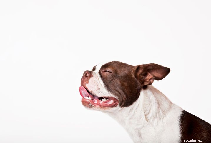
- Swelling of the neck under the jaw
- Swelling under tongue
- Swelling near the eye (rare)
- Trouble eating or swallowing
- Difficulty breathing (rare)
Sialoceles are generally painless unless they become large enough to put pressure on another part of the anatomy. Signs exhibited will depend on the type of sialocele.
Without treatment, sialoceles can become infected and abscessed. Contact your veterinarian if you notice any unusual swelling in the mouth or near the neck, jaw, or eye.
Causes of Sialoceles in Dogs
The exact cause of sialoceles is not known, but they are likely caused by traumatic injuries to the tissues of the salivary glands and ducts.
- Oral injury from chewing on an object
- Bite wounds from another animal
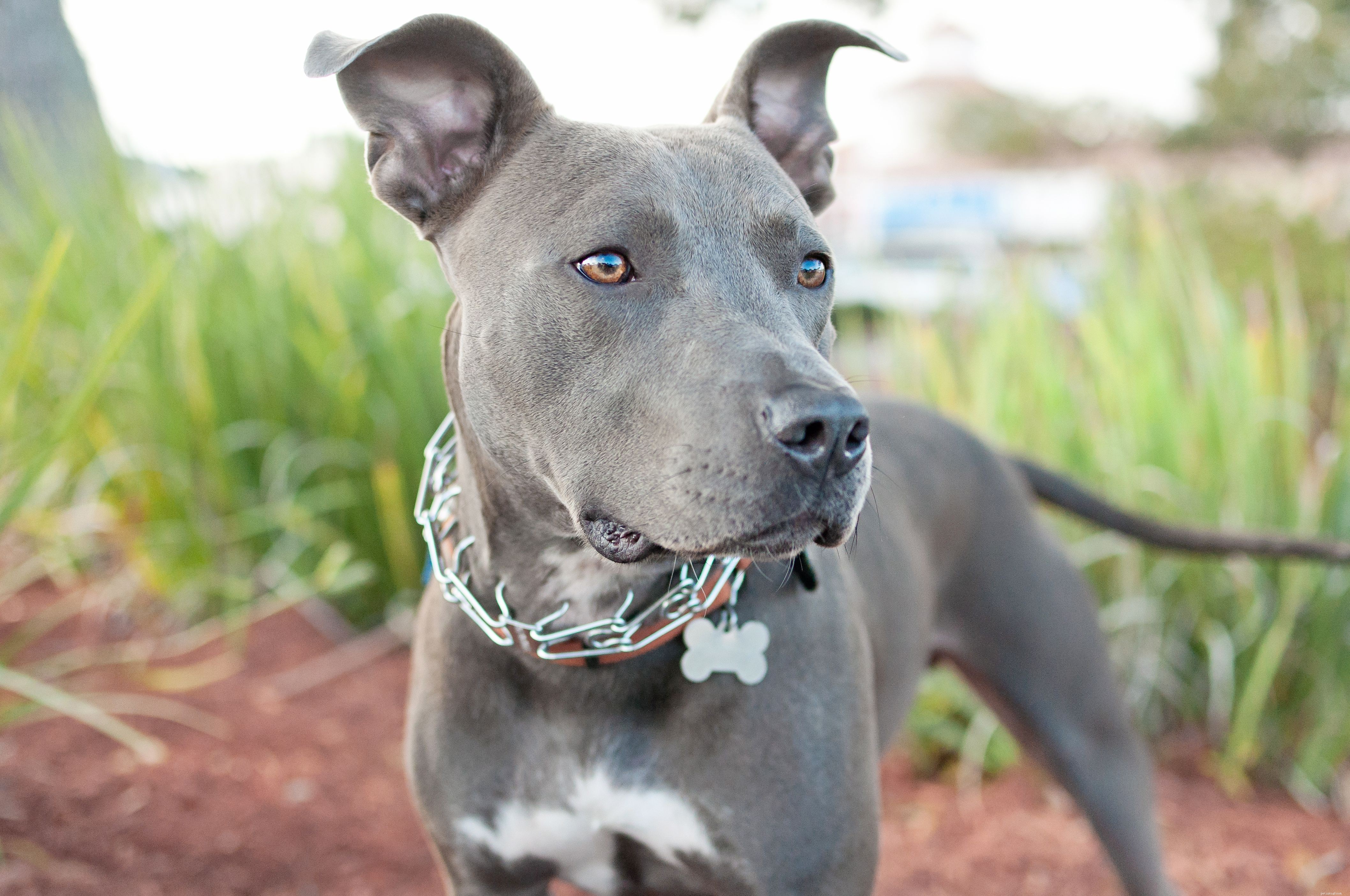
- Choke collar injury from pulling
Any dog breed can develop sialoceles, but German shepherds, dachshunds, poodles, and Australian silky terriers are more often affected.
Diagnosis
After discussing your pet's history, your veterinarian will perform a physical examination and look closely at the swollen area. Your vet may want to aspirate the area with a needle and syringe to collect the fluid for testing. Your dog may need sedation for this depending on the location.
The fluid from a sialocele is generally clear, yellowish, or blood-tinged in color and slightly viscous like saliva. Your vet may be able to see right away that it is saliva, but will likely send the fluid to a lab for analysis to be certain. A veterinary pathologist will analyze the fluid to determine what kinds of cells are present and confirm whether or not the swelling is a sialocele. This analysis can also rule out infections, cancer, and other potential causes for the swelling.
Treatment
Sialoceles typically require surgical intervention. In some cases, a sialocele can be drained to offer temporary relief until surgery can be performed. Most sialoceles will eventually recur after being drained. Continued draining is not recommended as it can lead to inflammation or infection.
Definitive treatment of sialoceles involves surgical removal of the affected salivary glands and associated ducts. This is a delicate procedure that is typically performed by a board-certified veterinary surgeon. Drains may be temporarily placed at the surgical site to prevent new fluid accumulation.
Most dogs recover well from salivary gland removal surgery with basic home care; complications are rare. Follow your veterinarian's recommendations for post-operative care. Give medications as directed. Keep the incision, drain sites, and any bandages clean and dry. Bring your dog back to the vet for follow-up visits as necessary.
How to Prevent Sialoceles in Dogs
Sialoceles are rare, but dog owners can still take steps to prevent injuries that may lead to sialoceles. Avoid using choke collars on your dog. Train your dog to walk on a loose leash to prevent injuries from pulling. Supervise your dog when gnawing on chews and toys, Keep your dog from chewing on sticks or other foreign objects.
Contact your veterinarian if you notice an injury to your dog's mouth or neck. Treatment of a fresh injury may prevent the development of a sialocele.
- Understanding and Preventing Bordetella Infections in Dogs
- Vitiligo in Dogs: Causes, Symptoms & Care Guide
- Leptospirosis in Dogs: Causes, Symptoms, and Human Health Risks
- Meningitis in Dogs: Key Symptoms, Causes, and Breeds at Risk
- Tetanus in Dogs: Symptoms, Causes, and Prevention Strategies
- Understanding Osteosarcoma in Dogs: Symptoms, Diagnosis, and Care
- Cheyletiella Mites in Dogs: Symptoms, Treatment, and Prevention